Optos Optomap® Dilation-Free Retinal Imaging
Why Optomap® Ultra-Widefield Retinal Imaging? Your eyes are the window to your health! Your retina (located in the back of your eye) is the only place in the body where blood vessels can be seen directly. This means that in addition to eye conditions, signs of other diseases (for example, stroke, heart disease, hypertension, and diabetes) can also be seen in the retina. Early signs of these conditions can show on your retina long before you notice any changes to your vision or feel pain. While eye exams generally include a look at the front of the eye to evaluate health and prescription changes, a thorough screening of the retina is critical to verify that your eye is healthy.
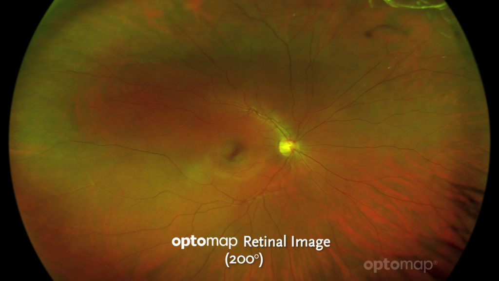
Getting an Optomap® image is fast, painless, and comfortable. Nothing touches your eye at any time. It is suitable for the whole family. To have the exam, you simply look into the device one eye at a time (like looking through a keyhole) and you will see a flash of light to let you know the image of your retina has been taken. Under normal circumstances, dilation drops might not be necessary, but your eye care practitioner will decide if your pupils need to be dilated depending on the health of your eyes. The image capture takes less than a half-second and they are available immediately for you to see your own retina. You see exactly what your eye care practitioner sees - even in a 3D animation.
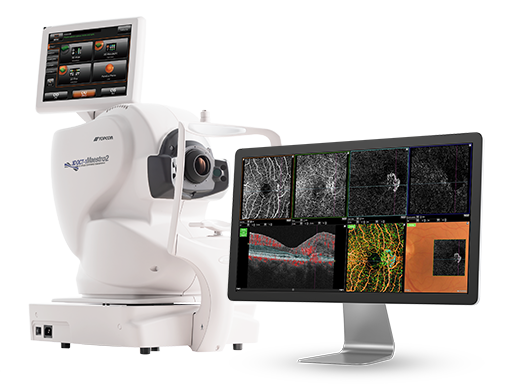
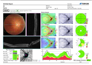
Maestro2® OCT
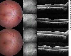
Optical coherence tomography (OCT) is a noninvasive imaging method that eye care specialists use to produce cross-sectional images of the deepest layers of the back part of your eye. It works by measuring wavelengths of light that reflect off the back of your eye (your retina). OCT allows eye specialists to examine your eye in layers and to measure the depth of important structures. This technology produces images akin to histological sections, providing optometrists with a virtual biopsy of the retina. Sometimes, providers use OCT to look at the front of your eye, too. It can help them diagnose defects in the front of your eye or plan for eye surgery. This level of detail enables early detection and intervention, potentially preventing vision loss or deterioration. The captured images are analyzed to detect abnormalities, retinal diseases, and related conditions that may affect your vision including:
- Bull’s eye maculopathy
- Central serous retinopathy
- Cystoid macular edema
- Diabetes-related macular edema
- Eye cancer
- Glaucoma
- Macular degeneration
- Macular holes
- Optic atrophy
- Posterior vitreous detachment (PVD)
- Retinal detachment
- Retinal tears
- Retinoschisis
The OCT scanner will scan one eye at a time. You might see a red line while it’s scanning. You won’t feel anything, and nothing will touch your eyes. The scan will take less than 1 minute. Since there is no radiation, there aren’t any risks or side effects associated with an optical coherence tomography.
RadiusXR Virtual Reality “VR” Perimetry
The area that you can see at one time without moving your eyes is called your field of vision or visual field. It may seem quite basic, but what you are able to see above and below a central point, and to either of its sides gives important clues into detecting many sight-threatening eye diseases, including and most commonly glaucoma. In early to moderate stages of glaucoma, many patients permanently lose peripheral vision without realizing it which makes visual field testing one of the most important tests during your annual eye exam. Aside from glaucoma, visual field testing can predict:
- Artery occlusion
- Brain tumors
- Multiple sclerosis
- Optic neuropathy
- Pituitary adenomas
- Retinitis pigmentosa
- Stroke
- Toxicity after taking medications including Plaquenil hydroxychloroquine
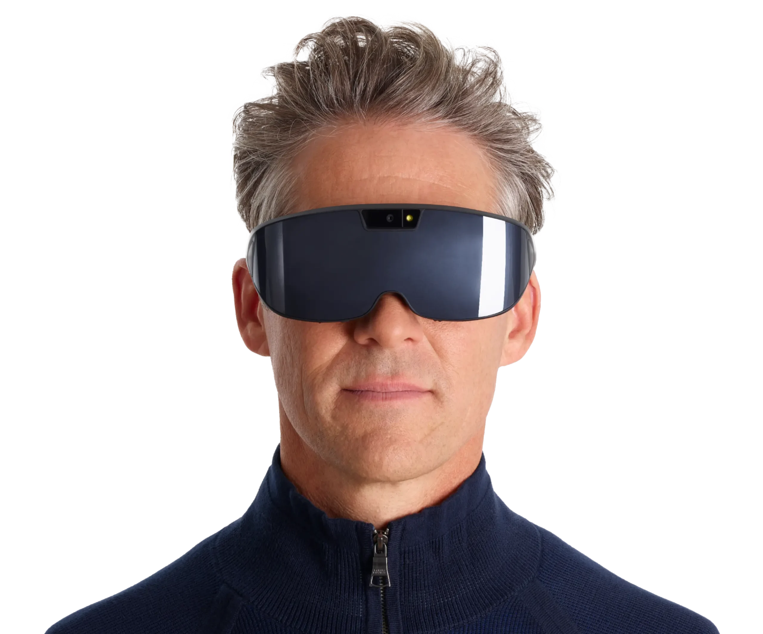
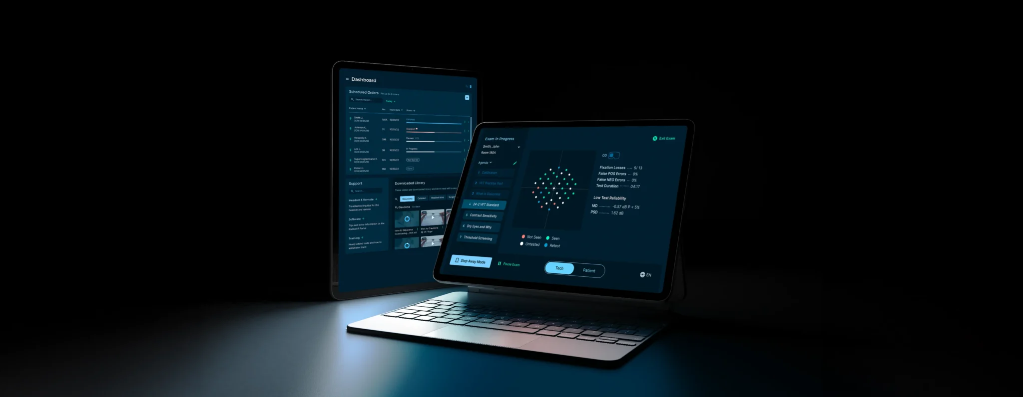
Our Virtual Reality VR headset technology brings visual field testing to a whole new level. Weighing a mere six ounces and fitting seamlessly like a pair of sunglasses, our VR headset allows the patient to adopt a more natural seated position and test both eyes simultaneously improving comfort and reducing test time. RadiusXR technology offers the most sensitive testing in clinical standards enabling more efficient detection of eye disease and better management and treatment of sight-threatening conditions.